Hard women's games. Foreign objects in the uterus Objects in the genitals
Hello Julia!
If you are sure that a small particle of an object broke off during masturbation, and not earlier, and remained in the genital tract, you should make every effort to remove the foreign body.
It is impossible to predict how a foreign body will behave in your body. Once in the vagina, a piece of plastic may not cause any disorders or pathological symptoms for a long time, it may come out with natural secretions, or, conversely, go deep into the uterus and further, with the most unpleasant consequences for health.
In some cases, you can remove a foreign object from the vagina yourself using your fingers, irrigation, or forceps. But this is only if the foreign body is shallow in the vagina and is determined by palpation. It all depends on where the foreign body initially entered, whether it caused damage to the epithelium and mucous surfaces, as well as on the size and shape of the foreign body.
In difficult cases, specialist assistance may be required to remove the object and prevent infections.
The entry of foreign objects into the genital tract is dangerous due to the possibility of developing bacterial infections, inflammation, and changes in the natural microflora.
The presence of a foreign body in the vagina can be manifested by symptoms of vulvovaginitis - leucorrhoea, hyperemia of the vestibule, burning, and spotting.
Carefully observe your condition and the nature of your discharge. You should be alerted to discharge with an unpleasant odor or unusual color. Sometimes the presence of a foreign body can cause vaginal bleeding. In this case, you should immediately consult a doctor.
If a foreign body remains in the vagina for a long time, erosion may develop. Sharp objects can lead to tissue perforation and secondary infection.
Diagnosis of a foreign body depends on the length of time the object is in the vagina, and includes a gynecological examination, colposcopy, vaginal probing, and smear examination.
If a foreign body is suspected of migrating from the vagina, the doctor may use a CT scan, scanogram, or abdominal x-ray to make a diagnosis. Ultrasound echography may also be needed to determine the location of a foreign body in the vagina and pelvis.
Treatment consists of removing the detected object using a finger, irrigation, instruments, or surgery.
The simplest and most effective way to remove a foreign body is to remove it with urethral forceps or tweezers after visualization using mirrors.
All symptoms of infection, pain and discharge usually resolve quickly after the foreign body is removed.
In case of prolonged presence of foreign objects in the genital tract and the formation of an infection, antibiotics may be prescribed.
The most serious complications arise when the infection penetrates into the deep pelvic tissues or peritoneal cavity.
Therefore, it is better not to risk your health and seek the help of a gynecologist.
This project was led by a 25-year-old woman. She has never given birth and has no history of STDs. Each photograph was taken at approximately 10:00 p.m., starting on the first day of the menstrual cycle. Throughout this project, she used condoms as a contraceptive method, and also to prevent seminal fluid during the photo shoot. She did not use tampons during her period.
This cycle is 33 days, which is the norm. The follicular phase of her cycle lasts until about 20–21 days. Favorable days for fertilization last several days from the 13th to the 21st day with ovulation on the 20th day. The luteal phase is 13 days (12–16 days is normal).
The above is for this cycle. As you can see, after ovulation on about the 20th day, her temperature began to rise due to increased progesterone, which in turn is produced by the corpus luteum. This temperature shift means that ovulation has already occurred.
She also monitored the position of the cervix throughout the entire cycle. Since the photo does not show whether the cervix is hard or soft, high or low. All this is clearly noticeable upon independent palpation. The uterus is tilted back (retroflexion), you may notice in several photos that the cervix is pointing upward. These are anatomical changes that are present in 20–30% of women, and most often a genetic trait.

The first day
The blood is red, there are slight cramps in the lower abdomen.
Breasts are slightly swollen.
Feelings are very sexual.

Second day
The blood is dark red.
The breast is normal.

Day three
Blood is brown, sometimes watery dark red.

Day four
Note the fresh blood.

Day five
Brown color.
Tired state.

Day six
Very light brown discharge.

Day seven
The neck is in a low, closed position.
There is sticky liquid on the neck.
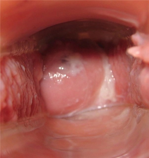
Day eight
The neck is low and closed.
Cervical fluid is white and sticky.

Day nine
The neck is low and closed.
Feeling dry.

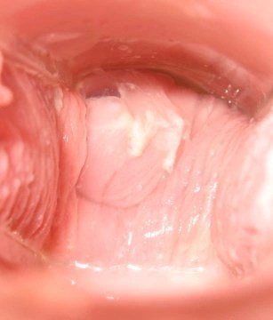
Day ten
The neck is low and closed.
Note the drop of blood and brown lump near the cervix (right). Perhaps from a stormy conversation on the same day, but later an endometrial polyp was diagnosed.

Day eleven
Cervical fluid is creamy.

Day twelve
Cervical fluid is white milky. Feeling wet.
I feel especially sexy.

Day thirteen
Copious watery discharge.
The neck is softened and moves upward.

Day fourteen
White, transparent, watery cervical fluid that stains underwear.

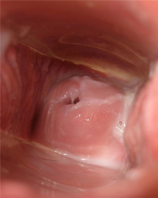
Day fifteen
The cervical fluid changes to a discharge that resembles egg white.
The neck is soft, open and high.

Day sixteen
Cervical fluid in the form of egg whites, very wet.
The neck is soft and high.


Day seventeen
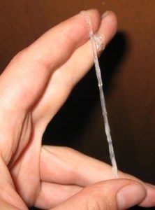
Cervical fluid is very thin, with whitish-yellow streaks. Sensual breasts, but not painful.
The liquid stretches between the fingers when stretched.
Day eighteen
Egg white.

Day nineteen
Egg white with a white tint.

Day twentieth
Minor back pain and cramps on the left side.
Suspicion of ovulation.
Feeling of strong sexuality.
Cervical fluid like gelatinous egg white.

Twenty first day
Cervical fluid is like glue.
The nipples are very sensitive and painful.

Day twenty two
Painful nipples.
The neck is in the middle position and slightly open.
Basal body temperature begins to rise.

Day twenty-three
Very sensitive nipples.
Feeling dry.

Day twenty-four
Very sensitive nipples.
Dry.
The neck is firm and high.

Day twenty five
Headache and fatigue.
Cervical fluid is dry/sticky.
Day twenty six
Breasts are swollen.
Basal body temperature is now noticeably higher, by about 1 degree.

Day twenty seven
Painful nipples, swollen breasts.
Cervical fluid is sticky.

Day twenty eight
Feeling dry.

Day twenty nine
Feeling dry.

Day thirty
Feeling dry.
The chest is heavy.

Day thirty one
Feeling bloated.
Dry, (note, fresh blood, a sign of impending menstruation).
Feeling of emotional instability.

Day thirty two
Light brown spots.
The neck is low and open.
Feeling tired.

Day thirty three
Pink spots.
Pain in the lower back.
Menstruation will begin tomorrow after waking up, 13 days after ovulation.
The article was taken from the internet! Who is not interested, don’t fu... fuck!
- a foreign object that has entered the lumen of the vaginal tube through the genital slit. The presence of a foreign body in the vagina is manifested by symptoms of vulvovaginitis - leucorrhoea, hyperemia of the vestibule, burning, and spotting. When an object is left in the vagina for a long time, bedsores, tissue necrosis, and ulcerative processes can develop. Diagnosis of a foreign body in the vagina includes a gynecological examination (vaginal or rectal), colposcopy, vaginal probing, and smear examination. Treatment consists of removing the detected object using a finger, irrigation, instruments, or surgery.

General information
Among the foreign bodies of the vagina that gynecology encounters in its practice, there may be objects that have been introduced accidentally or as a result of injuries to the external genitalia, as well as those introduced specifically (for the purpose of sexual satisfaction, contraception, termination of pregnancy, medical procedures).
In girls, lumps of baby powder, grains of sand, pins, threads, small parts of toys, buttons, tacks, etc., may be found in the vagina, accidentally or during play or pranks. During puberty, various foreign bodies are more often introduced into the vagina during masturbation. Foreign objects enter the vagina of sexually mature women during masturbation, attempts to prevent pregnancy or an attempt to cause spontaneous abortion, and also, much less frequently, during gynecological manipulations.
Symptoms of a foreign body in the vagina
Once in the vagina, a foreign body may not cause any disorders or pathological symptoms for a long time. Subsequently, prolonged irritation of the mucous membrane or the addition of a microbial infection is accompanied by symptoms of colpitis (in girls - vulvovaginitis): swelling of the vulva, hyperemia of the vestibule, liquid and milky whites or mucopurulent discharge, sometimes with the presence of blood and a sharp putrid odor. Heavy discharge often causes maceration of the perineal skin and pyoderma.
Ascending infection can lead to the development of cervicitis, endometritis, urethritis, cystitis and intra-abdominal infection, adhesions in the pelvis, and infertility. If sharp-pointed objects are in the vagina, damage to the vaginal wall may occur with the development of bleeding, requiring emergency care. In severe cases, penetrating wounds of the vaginal wall with injuries to the bladder, rectum, and pelvic organs are possible. When a foreign body remains in the vagina for a long time, bedsores, tissue necrosis and ulceration, vaginal stenosis, genitourinary and vaginal-rectal fistulas can develop.
Diagnosis of a foreign body in the vagina
The presence of foreign bodies in the vagina is recognized during gynecological examination, examination with speculum and colposcopy. For virgins, a bimanual recto-abdominal examination, vaginal probing and vaginoscopy are performed. In cases of vulvovaginitis, a bacteriological examination of the smear is necessary.
Long-term, persistent and treatment-resistant vulvovaginitis in children always causes the gynecologist to assume that there is a foreign body in the vagina. If migration of a foreign body from the vagina is suspected, a survey of the abdominal cavity, ultrasound, or CT may be required. Carrying out diagnostics allows you to find out the nature of the foreign body in the vagina and choose the right method for its removal.
Removing a foreign body from the vagina
A difficult task when removing a foreign body is the need to avoid injury to the vaginal walls and, if the hymen is intact, damage to its integrity. In typical cases, a foreign object can be removed from the vagina with a finger, urethral forceps, tweezers or a clamp with long narrow jaws, or a Volkmann spoon. If smooth, round objects (balls, beads, buttons, etc.) enter the vagina, the foreign body can be pushed out with the help of a finger inserted into the rectum and a helping hand through the abdominal wall.
To remove fibrous tissue (wool, threads), the preferred method is vaginal lavage. If there is a suspicion of injury to the vagina, as well as detection of traumatic objects, the foreign body is removed in the operating room. The presence of vaginal stenosis, which prevents the removal of a foreign body, requires dissection of the scars. In severe cases, vaginoplasty is necessary after removal of the foreign body. In the future, to relieve symptoms of vaginitis, vaginal douching and antibiotic therapy are carried out.
Prevention of foreign bodies entering the vagina
Preventing foreign bodies from entering the vagina comes down to careful monitoring of children, their games and hygiene, and proper upbringing of girls in families and children's groups. The use of objects not intended for insertion into the vagina must be carried out strictly for their intended purpose, and vaginal hygiene and contraceptive means - in accordance with the instructions. Also, in order to prevent foreign bodies from entering the vagina, careful gynecological manipulations are necessary.
A foreign object can be located both in the uterine cavity itself and in its wall. Such a neighborhood is manifested by specific symptoms. The presence of a foreign body in the uterus is always accompanied by a whole complex of pathological changes. Firstly, a foreign object will cause bleeding. It can also cause an inflammatory process. In this case, the woman may have chronic endometritis.
Such changes are almost always accompanied by pain, so a woman may come to see a doctor complaining of pain in the lower abdomen. Due to the presence of a foreign object in the uterus, secondary infertility may develop.
Based on complaints and external examination, it is not possible to make a final diagnosis. Hysteroscopy can help detect a foreign object in the uterus; ultrasound examination and sounding will be slightly less useful.
The only method of treatment is mandatory removal of the foreign object. A long stay of a foreign object inside the reproductive organ can become a source of serious problems. A foreign body in the uterus is always a time bomb.
According to statistics, threads, plastic hangers and parts of the IUD are most often found. All these are remnants of intrauterine contraceptives, surgical ligatures. Very rarely, fragments of bone tissue left from the fetus after an abortion act as a foreign body.
These parts can be found in patients who have had an abortion in the past, especially after fourteen weeks. If a foreign object in the uterus is encapsulated, then there will be no picture corresponding to the fact that there is something in the cavity. In this case, the foreign body will not be detected at all.
Since the uterus is a muscular organ, it can contract. If a foreign object is located freely in the uterus, then during contractions it will change its location.
A foreign body easily injures the walls of the uterus. The hole can be complete when the foreign object extends beyond the walls of the organ and incomplete when the foreign body stops at the level of the myometrium. In this case, the object from the uterus is able to move to another place.
An infectious process that can develop due to the presence of a foreign body can also pose a danger. It is very difficult to cure such inflammation, especially if the source of infection continues to remain in the uterus. Most often, the cause of endometritis is lavsan or silk ligatures, which remain after various medical procedures, namely cesarean section, conservative myomectomy.
In order to understand the seriousness of the situation, it is enough to consider a simple example. Silk ligatures remained in the uterine cavity after the operation. Silk is a material that does not dissolve over time.
After some time, purulent discharge begins to appear. The period between the entry of the ligature into the uterus and the onset of suppuration can be very short. If, after the first signs of inflammation appear, a woman is prescribed antibiotics, there will be no significant improvement in the woman’s condition.
Symptoms
A foreign body in the uterus can affect your period. Most often, the discharge during menstruation in this case becomes abundant and prolonged. Blood loss in this case may exceed the normal level several times. Very often, menstruation is preceded by purulent leucorrhoea. In the period between menstruation, discharge with bloody clots may appear.
Pain
Severe pain in the lower abdomen also appears when there is a hole in the wall of the uterus. An incorrectly installed IUD can injure the uterus. At the same time as the uterus, other pelvic organs can be injured. A foreign object located in the uterus can subsequently be found in the bladder, in the area of the uterine appendages and appendix.
Bleeding
A foreign body can cause severe bleeding. The main symptom of this pathology is bloody discharge from the vagina. A woman may feel a bout of weakness and dizziness. With severe bleeding, an increase in signs of anemia is observed.
Diagnostics
Hysteroscopy allows you to accurately determine the location of the foreign body. Other examination methods are less accurate and can be used as auxiliary ones. For example, with the help of a probe a foreign body can be detected, but it will not be possible to find out where it is in this way. It’s impossible to determine its shape and size.
Hysteroscopy also allows you to determine how a foreign object is located in relation to the walls of the uterus and other pelvic organs. Ultrasound examination can help in detecting the remains of intrauterine contraceptives.
These fragments can be found both in the uterine cavity and inside the myometrium. Using ultrasound, it is almost impossible to detect bone remains left after an abortion and ligature. Foreign objects in the uterus can also be detected using radiography, but many of the nuances of finding a foreign body in the uterus may be missed during this examination.
Hysteroscopy
What the doctor sees during hysteroscopy depends on the type of object that is in the cavity. If a foreign object was in the uterus for quite a long time, then it could well be overgrown with endometrium. If a foreign object is tightly adjacent to the wall of the uterus, then a bedsore may form in this place. The hysteroscope allows you to carefully remove this object.
During hysteroscopy, a foreign body is removed using a special instrument through the hysterocope channel. Removing a foreign body without proper supervision is not recommended as the foreign body may disintegrate when removed.
When removed, a deformed foreign object can injure the walls of the uterus. If possible, it is better to get a foreign body during the period when the corpus luteum is maturing. This period occurs approximately in the middle of the cycle. If, in the opinion of doctors, there is a threat to the woman’s life, then the foreign object is removed immediately.
Foreign objects in the cervix may appear after medical procedures. But in practice, a foreign body that has entered the cervix is found not in the cervix itself, but in the fornix.
Complications
If there is perforation of the uterus and if severe inflammation is associated with this injury, conservative treatment may not be effective. In this case, doctors may resort to removing the uterus.
Next, the patient is prescribed intensive antimicrobial treatment. When a foreign body is detected, special attention should be paid to the condition of the uterus. After artificial termination of pregnancy, removal of the intrauterine device, it is recommended to conduct an inspection of the uterine cavity
Foreign objects should not be left in the uterus. A common phenomenon accompanying the presence of a foreign body in the uterus is pyometra. This is the name for a condition in which the uterus is filled with purulent contents. This complication is difficult to treat, even with the latest generation of antibiotics.
The most severe complication of this pathology is necrosis of uterine tissue. Suppuration can cause sepsis. All these conditions pose a threat to a woman’s life. After removing foreign objects from the uterus, the effectiveness of certain drugs is checked before prescribing antibiotic therapy.
 How much fabric is needed for a dress: simple methods and basic calculation nuances
 Beautiful Christmas tree decorations made of plywood: we make decorations with our own hands using a jigsaw
 Manicure with acrylic paints at home