Polymorphism of feelings: how skin receptors work. Types of skin receptors Krause flasks perceive

Skin receptors are responsible for our ability to feel touch, heat, cold and pain. Receptors are modified nerve endings that can be either free non-specialized or encapsulated complex structures that are responsible for a certain type of sensitivity. Receptors perform a signaling role, so they are necessary for a person to effectively and safely interact with the external environment.
The main types of skin receptors and their functions
All types of receptors can be divided into three groups. The first group of receptors is responsible for tactile sensitivity. These include the bodies of Pacini, Meissner, Merkel and Ruffini. The second group is
thermoreceptors: Krause flasks and free nerve endings. The third group includes pain receptors.
The palms and fingers are more sensitive to vibration: due to the large number of Pacini receptors in these areas.
All types of receptors have different zones in terms of the width of sensitivity, depending on the function they perform.
Skin receptors:
. skin receptors responsible for tactile sensitivity;
. skin receptors that respond to changes in temperature;
. nociceptors: skin receptors responsible for pain sensitivity.
Skin receptors responsible for touch sensitivity
There are several types of receptors responsible for tactile sensations:
. Pacinian corpuscles are receptors that quickly adapt to changes in pressure and have wide receptive fields. These receptors are located in the subcutaneous fat and are responsible for gross sensitivity;
. Meissner's bodies are located in the dermis and have narrow reception fields, which determines their perception of fine sensitivity;
. Merkel bodies - slowly adapt and have narrow receptor fields, and therefore their main function is to sense the structure of the surface;
. Ruffini's bodies are responsible for the sensation of constant pressure and are located mainly in the area of \u200b\u200bthe soles of the feet.
Also, receptors located inside the hair follicle are separately isolated, which signal the deviation of the hair from its original position.
Skin receptors that respond to changes in temperature
According to some theories, there are different types of receptors for the perception of heat and cold. Krause flasks are responsible for the perception of cold, and free nerve endings are responsible for the perception of hot. Other theories of thermoreception claim that it is the free nerve endings that are designed to sense temperature. In this case, thermal stimuli are analyzed by deep nerve fibers, while cold stimuli are analyzed by superficial ones. Between themselves, the temperature sensitivity receptors form a "mosaic" consisting of cold and heat spots.
Nociceptors: skin receptors responsible for pain sensitivity
At this stage, there is no final opinion regarding the presence or absence of pain receptors. Some theories are based on the fact that free nerve endings, which are located in the skin, are responsible for the perception of pain.
Prolonged and strong pain stimulation stimulates the emergence of a stream of outgoing impulses, and therefore, adaptation to pain slows down.
Other theories deny the presence of separate nociceptors. It is assumed that tactile and temperature receptors have a certain threshold of irritation, above which pain occurs.
Table of contents of the subject "Sensory Systems. General Physiology of Sensory Systems. Classification of Receptors. Somatovisceral Sensory System.":1. Sensory systems. Sense organs. Physiology of the sense organs. Functions of sensory systems. Sensory perception. Stages of sensory perception.
2. General physiology of sensory systems. Classification of receptors. appropriate receptors. Mechanoreceptors. Chemoreceptors. Photoreceptors. Thermoreceptors.
3. Classification of receptors. Monomodal and polymodal receptors. Nociceptors (pain receptors). Exteroreceptors. Interoreceptors.
4. Conversion of stimulus energy in receptors. receptor potential. Absolute threshold. The duration of the sensation. Receptor adaptation.
5. Receptive fields. receptive field. The magnitude of the receptive fields. Types of receptor fields.
6. Processing (processing) of information in the switching nuclei and pathways of the sensory system. Lateral inhibition.
7. Downward inhibition (gain). Negative feedback mechanism. positive feedback mechanism. Multichannel.
8. Subjective sensory perception. The absolute threshold of sensation. differential threshold. Discrimination threshold. Weber's law. Weber-Fechner law. Stevens scale.
9. Somatovisceral sensory system. somatovisceral system. Tactile sensitivity. Taurus Pacini, Meissner, Ruffini. Merkel discs. Tactile corpuscles of Pinkus-Iggo. Krause flasks.
10. Proprioceptive sensitivity. proprioceptive sensations. Proprioceptors. proprioceptive perception.
Somatovisceral sensory system. somatovisceral system. Tactile sensitivity. Taurus Pacini, Meissner, Ruffini. Merkel discs. Tactile corpuscles of Pinkus-Iggo. Krause flasks.
Somatovisceral sensory system
Somatovisceral system processes sensory information of several modalities, receiving it from the skin, muscles, joints, internal organs and blood vessels. It contains different types of receptors: mechanoreceptors, thermoreceptors, chemoreceptors, osmoreceptors, nociceptors. The incoming information is transmitted along afferent fibers as part of sensory and mixed nerves, processed by separate pathways and switching nuclei, and then delivered to the somatosensory cortex, which occupies the postcentral gyrus of the brain and is organized topically.
Tactile sensitivity
Part somatovisceral system, which provides a sense of touch, includes several types of skin mechanoreceptors, represented by free nerve endings or encapsulated, i.e., encapsulated from connective tissue or modified cells of the epidermis (Fig. 17.4). Free nerve endings innervate the hair follicles of vellus hair covering most of the human body, as well as coarse hair growing on the head, in the armpits, on the pubis, and in men also on the face. The free nerve endings of the hair follicles are mechanoreceptors and are excited when the hair is displaced or twitched. Another variety of free nerve endings is found in the epidermis and in the papillary dermis, most of them are nociceptors or thermoreceptors, but some belong to mechanoreceptors that specifically respond to weak near-threshold irritation. It is assumed that when this type of receptor is irritated, sensations of tickling and itching occur.
Rice. 17.4. Scheme of distribution of mechanoreceptors in human skin. Free nerve endings have a high threshold of irritation and respond poorly to changes in stimulus intensity. Rapidly adapting receptors (Pacini corpuscles, hair follicle receptors) serve as sensors for the speed of acting stimuli, and slowly adapting receptors (Merkel's discs, Ruffini corpuscles) serve as sensors for the intensity of the acting stimulus. The presence of several varieties of receptors makes it possible to transmit afferent signals about different properties of the same stimulus.Among encapsulated endings distinguish Pacini corpuscles, Meissner, Ruffini, Merkel discs, tactile Pinkus-Iggo bodies, Krause flasks. Depending on the structure and shape of the capsule, the nerve endings are subject to the strongest influence either as a result of pressure from a perpendicular stimulus, or as a result of the lateral displacement of the capsule, which plays the role of a mechanical energy converter of external stimuli. Most of the encapsulated receptors are found in the hairless skin of the fingers and toes, palms and soles, face, lips, tongue, nipples, and genitals, where they are distributed in varying densities and depths. Pacinian corpuscles are also found in tendons, ligaments, and mesentery.
skin mechanoreceptors differ in the rate of adaptation to the acting stimulus. Rapidly adapting (phasic) receptors are excited only at the moment of displacement of the skin and hair and serve as sensors of the speed of the stimulus. This property is inherent Meissner corpuscles, hair follicle receptors, and especially Pacinian corpuscles, capable of responding to changes in the speed of a continuing stimulus. Slowly adapting (tonic) receptors do not stop generating action potentials during prolonged action of the stimulus if it puts pressure on the skin: such receptors serve as sensors for the intensity of the current stimulus (Ruffini bodies, Merkel discs).
Table 17.1. Spatial two-point threshold in different parts of the body
Area of receptive fields of sensory neurons, innervating Meissner bodies And Merkel discs, averages about 12 mm2, and in neurons with endings in the form Pacini's corpuscle And Ruffini she's much bigger. receptive fields sensory neurons that differ in their receptors overlap, therefore, when a complex of stimuli acts on the skin, different types of receptors are simultaneously excited, which makes it possible to feel all the dynamic and static properties of such a complex. The processing and analysis of information from signals from various receptors occurs at the highest levels of the sensory system, which form a complex perception of stimuli acting on the surface of the body. The density of mechanoreceptors in different parts of the skin is not the same, which determines different indicators of spatial differential threshold, i.e., the smallest distance between two points, the irritation of each of which is felt separately (Table 17.1). The data in the table should not be taken as a reference, since differential sensitivity varies from person to person.
Encapsulated receptors are innervated myelinated fibers of primary sensory neurons that conduct nerve impulses to the CNS at a speed of about 30-70 m/s. Unmyelinated fibers transmit action potentials from free nerve endings at a much lower speed - about 1 m / s, so the sensation of the stimulus acting on them occurs relatively later. The central processes of primary sensory neurons enter the spinal cord as part of the posterior roots and are divided into collaterals in the posterior horns of the spinal cord. Ascending collaterals reach the switching nuclei of the posterior column of the medulla oblongata, from where specific information is transmitted to the opposite side of the brain along the lemniscal pathway, goes to the projection nuclei of the thalamus, and then to the somatosensory cortex, with the participation of which the sense of touch is formed.
Krause bulbs) encapsulated nerve endings, consisting of terminal branches of a sensitive nerve fiber, an internal glial bulb and an external connective tissue capsule; located in the connective tissue basis of the mucous membranes, under the epidermis and among the muscle fibers of the tongue.
1. Small medical encyclopedia. - M.: Medical Encyclopedia. 1991-96 2. First aid. - M.: Great Russian Encyclopedia. 1994 3. Encyclopedic dictionary of medical terms. - M.: Soviet Encyclopedia. - 1982-1984.
See what "Krause flasks" are in other dictionaries:
- (W. J. F. Krause, 1833 1910, German anatomist; syn. Krause bulbs) encapsulated nerve endings, consisting of terminal branches of a sensitive nerve fiber, an internal glial flask and an external connective tissue capsule; ... ... Big Medical Dictionary
- (W. J. F. Krause, 1833 1910, German anatomist) see Krause flasks ... Big Medical Dictionary
- (organs of touch). Although the sense of touch is undoubtedly characteristic of all animals, we find completely separate O. organs only in animals that are relatively highly organized. Not to mention the protozoa (Protozoa) and sponges (Porifera), which are what ... ... Encyclopedic Dictionary F.A. Brockhaus and I.A. Efron
- (W.J.F. Krause, 1833 1910, German anatomist) see Krause flasks ... Medical Encyclopedia
Receptors located in the outside. integument, muscles, tendons, joints, fascia, some mucous membranes (lips, tongue, genitals); perceive mechanical action. (touch, pressure), temperature and pain stimuli. Among… … Biological encyclopedic dictionary
Special perceiving devices Receptors embedded in the skin, musculoskeletal system (muscles, tendons, joints, etc.), some mucous membranes (lips, tongue, etc.). With the help of O. o. the body perceives complex ... ...
skin nerves- The skin contains blood vessels and sensory, motor, vasomotor, sympathetic and secretory nerves. The endings of sensory nerves are located in the epidermis, thanks to them the perception of pain is carried out. Tactile… … Atlas of human anatomy
NERVE ENDINGS- NERVE ENDINGS, junctions of nerve fibers with elements of various tissues of the body. Formations that connect nerve cells with each other, the so-called. pericellular devices, can also be classified as N. o. (see Non-urgent theory) ...
A set of anatomical and physiological mechanisms that ensure the perception, analysis and synthesis of mechanical, thermal, chemical and other stimuli falling from the external environment on the skin and some mucous membranes (oral and nasal cavities, ... ... Great Soviet Encyclopedia
EIMER AUTHORITY- EIMER'S organ, feeling the nerve endings of the mole's snout. They are located in the epidermis and are cylindrical accumulations of epithelial cells, which include 1-3 thick torn fibers, curving between the cells, and a number of thin ... ... Big Medical Encyclopedia
Biophysics report:
“Mechanoreception”
1. Appointment
For active existence in the environment, higher organisms have a large number of analyzers specializing in different types of influence: on light (vision), on sound (hearing), on taste and smell (smell), on touch and temperature (touch), on gravity . Mechanoreception enters the sense of touch and, together with the vestibular apparatus, allows the body and consciousness to most fully represent the position of the limbs and the body in space.
2. Functions
The analyzer of mechanoreception, like any other analyzer, includes three spatial-functional parts: the receptor (receiving and transforming mechanical action into an electrical impulse), conduction pathways (impulse transmission) and the nerve center (analysis of the information received and the formation of an efferent response). All these parts provide:
Perception of mechanical effects on the skin: localization, direction of movement, speed of the deforming source, its vibrations (tactile reception);
- Perception of mechanical shifts within organs and muscles in order to determine the position of the limbs and body in space (proprioception);
- Perception of effects on the hairline.
- Transformation of a signal from mechanical to electrical, which can be transmitted through neurons.
- Transmission of impulses at high or low speed to the nerve center.
- Formation in the mind of a general picture of the position of the body and limbs in space.
- Providing the autonomic nervous system with information about the position of the body and its control (maintaining the vertical position of the body with a possible falling asleep in this position, information about oculomotor movements in a dream).
3. Device and principle of operation.
Typical mechanoreceptors, as a rule, are encapsulated formations. Some of them are called superficial end organs, since they are located superficially in the skin. These are Merkel's discs, Meissner's bodies, Vater-Pacini's bodies, Dogel's bodies, Krause's flasks, Ruffini's bodies, neurotendinous spindles, neuromuscular spindles and others (Fig. 1).
Fig.1. Different types of mechanoreceptors
Lamellar bodies (according to the old terminology of Vater-Pacini bodies) are located in the connective tissue of internal organs and deep layers of the skin, especially on the fingertips, on the mesentery, in the mammary gland, intestines and other internal organs. They have the appearance of rounded formations.
Structural components are:
The inner flask (bulb), formed by modified lemmocytes, into which nerve fibers penetrate;
- outer flask - a layered connective tissue capsule of fibroblasts and collagen fibers, forming concentric plates, between which there is a liquid.
The interior of the capsule contains flat, concentrically arranged neuroglial cells that delimit the inner flask. The receptor fiber enters the inner flask from one of its poles and forms contacts with glial cells. The terminal part of the receptor fiber contains small spherical mitochondria and light synaptic vesicles. The outer layer of the capsule consists of a powerful connective tissue membrane formed from flat sickle-shaped cells and connective tissue fibers, between which there is an interstitial fluid. Lamellar bodies perceive the sensation of vibration, tension, pressure on organs and intraorgan pressure. The principle of operation of Vater-Pacini bodies is poorly understood today. The connective tissue plates and the interstitial fluid of the capsule probably contribute to increased pressure on the nerve ending, as a result of which the axolemma is deformed, its permeability changes, and a potential is generated. It is believed that the site of the action potential in the little bodies of Pacini is the region of the first intercept of Ranvier.
Tactile bodies (Meissner) located in the papillary dermis, have an ellipsoidal shape and small size. These are tactile receptors that respond to touch. Present in the dermis of the skin, especially in the fingertips, soles, nipples, eyelids, lips, and genitals. In the center of the Meissner body is a spirally coiled, unmyelinated branching of the myelin fiber, which passes through transversely arranged oval cells resembling Schwann cells. Outside, the body is covered with a connective tissue capsule. The inner flask consists of glial cells lying perpendicular to the long axis of the body, between which there are branches of dendrites. Outside there is a very thin, layered capsule turning into perineuritis - the outer flask. A slight deformation of the capsule is transmitted to gliocytes and further to the dendrites.
Merkel cells located under the epidermis, have large irregular nuclei and microvilli extending to the epidermal cells. At their bases are disc-shaped endings of sensory axons (Merkel's discs). A group of 10 - 20 Merkel cells form synaptic contacts with the endings of one sensory axon (Pinkus-Iggo bodies). Merkel cells respond to sudden movements of the skin, such as when stroked.
Ruffini's bodies lie in the connective tissue of the skin and joint capsules: they perceive pressure and look like spindle-shaped structures. The inner flask is formed by glial cells between which there are terminals of dendrites with extensions at the ends. The capsule is well defined.
Krause flasks- small round bodies, which are mechanoreceptors and cold receptors. They lie in the dermis of the skin, the mucous membrane of the oral cavity, subglottis, in the conjunctiva of the eye. The inner flask is formed by flat gliocytes between which thin branches of the dendrite form plexuses in the form of a glomerulus. The outer capsule is very thin.
Dogel's genital bodies- are located in particularly sensitive areas of the skin (external genitalia, mammary glands). They are similar in structure to Krause's flasks, but unlike them, several processes from neurocytes enter the body, which causes a strong irradiation of excitation. They respond to pressure with impulses that cause sexual arousal.
Mechanosensitive free endings in the skin. They are unmyelinated axons, they do not have corpuscular structures. These include hair follicle receptors that respond to pressure from hair movement.
Neuromuscular spindles- Stretch receptors of striated muscles - nerve endings that have both sensory and motor innervations. Sensitive nerve fibers in coils abundantly braid several muscle fibers, forming around them like a clutch. In this area, muscle fibers become thinner, the number of myofibrils in them decreases, and the number of nuclei increases sharply. Neuromuscular spindles are surrounded by a connective tissue capsule.
Motor nerve fibers form small neuromuscular synapses along the edges of intrafusal fibers, providing their tone, regulating the length of the fibers. All free space between muscle fibers is filled with fluid and is limited by a thin capsule. A change in muscle tone leads to a change in fluid pressure and is transmitted to the dendrites. The ring-spiral endings respond to changes in the length of the muscle fiber and to the rate of this change; The number of spindles in a muscle depends on its function and the higher, the more precise movements it has.
Nerve-tendon spindles - stretch receptors, located at the junction of the muscle with the tendon, spindle-shaped structures 0.5-1 mm long. Each spindle has a capsule of fibrocytes, which encloses a group of tendon bundles entwined with nerve fiber endings. Excitation of receptors occurs when the tendon is stretched during muscle contraction.
From the receptor, the impulse moves along the reflex arc through the spinal cord and brain stem to the nuclei of the thalamus and further to the cortex.
4. Performance
Number and placement density of receptors:
The total number of tactile receptors is estimated at 10 million pieces, grouped into 1 million afferents.
- Number of free afferent endings in the skin: 50% of the total number of skin afferents.
- Density of placement of free nerve endings in the skin: 170 pcs/cm2
Dimensions and other structural parameters:
Meissner bodies: 50-140 µm
- Krause flasks: 40-150 microns
- Ruffini bodies: 1-2 mm
- Vater-Pacini bodies: 0.5-5 mm
- Nerve tendon spindles:
- Dimensions: 0.5-1 mm
- Number of muscle fibers in the spindle: 15 pieces
- neuromuscular spindles
- Number of muscle fibers in the spindle: 1-8 pieces
- Diameter of outgoing nerve fibers: 17 µm (primary) and 8 µm (secondary)
Transmission speed:
Psychophysical assessment of the capacity of tactile channels: 5 bps
- The speed of transmission of impulses from free nerve endings: 1 m / s (C-fibers, type IV)
- Velocity of impulse transmission from encapsulated receptors: 50 m/s (A-fibers, type II)
Resolution and thresholds of sensations:
Threshold of sensation of skin receptors under pressure: 10 microns
- Simultaneous spatial threshold of skin receptors:
- On lips and fingertips: 1-3 mm
- On the back, shoulders, hips: 50-100 mm
- Sequential spatial threshold of skin receptors:
- On lips and fingertips: 1 mm
- On the back, shoulders, hips: 10-20 mm
- Threshold of vibration sensation: 150-300 Hz
- Minimum perceived vibration amplitude: 1 µm
Modality(specificity to influences):
Ruffini bodies and Krause flasks, in addition to mechanical effects, detect temperature, i.e. polymodal. And Meissner's bodies can feel the vibration. Non-encapsulated free afferents are also polymodal.
- Other receptors are unimodal.
Receptor fields(reception area):
For tactile discs, the area is equal to the anatomical dimensions.
- For Merkel's discs, this is an agglomeration of 30-50 discs served by one nerve fiber.
- For Vater-Pacchini bodies, this area is anatomically larger.
Adaptation(see Table 1).
Table 1. Classification of skin mechanoreceptors according to the rate of adaptation and adequate stimuli.

5. Regulation
Information from the receptors is transmitted to the central nervous system through the spinal or cranial nerves. Axon branches that carry sensory information form synaptic endings on neurons in the spinal cord (or brainstem in the case of cranial worms), then travel to the higher parts of the nervous system (stem nuclei, which in turn relay information above). In each of these parts of the nervous system, the flow of sensory information can be filtered - accentuated or, conversely, blocked.
Sensory systems are built on a hierarchical principle: signals from receptors enter the lower levels of the central nervous system (spinal cord or brain stem), from where they are transmitted to higher sections (thalamic nuclei, cerebral cortex, basal ganglia). At each of these successive stages, sensory information is transformed and filtered.
The flow of information is not one-way, as the higher departments of the hierarchy send signals to the lower departments. In addition, sensory information is not processed by a chain of sequential structures, but rather by many areas of the brain simultaneously (or, as they say, in parallel). Parallel processing of sensory information becomes evident in higher areas of the brain, such as the cerebral cortex. Here, separate areas specialize in the processing of individual elements of information.
Sensory information of various kinds is not processed separately. In many areas of the brain, called associative, there is a mixture of modalities - for example, in the parietal cortex and the colliculus. Neurons in these regions of the brain respond to various stimuli, such as visual, tactile, and auditory.
The structures that control sensory information are the cerebral cortex (in particular, the prefrontal cortex, which plays an important role in controlling attention), the basal ganglia, the reticular formation, the thalamus (in particular, the reticular nucleus of the thalamus), and other structures.
One of the main mechanisms for filtering sensory information is inhibition, which is produced by GABAergic synapses. As a rule, the neuron that transmits sensory information is excitatory. These excitatory signals are filtered by inhibitory neurons. Inhibition can be presynaptic (that is, blocking the transmission of signals along the sensory axon to any neuron) or postsynaptic (hyperpolarizing neuron that receives sensory signals). Postsynaptic inhibition makes it possible to selectively block signals, since the receiving cell remains able to respond to other, unblocked stimuli.
The filtering of sensory signals is also influenced by neurotransmitters such as acetylcholine, dopamine, endorphins, and others.
6. Energy dependence
Since the entire analyzer of mechanoreception is a derivative of the nervous tissue, the energy dependence is especially high. Neurons are supplied with nutrients and oxygen through the neuroglia and the blood vessels located in it. During hypoxia and blood clots, the nutrition of individual areas (for example, skin or organs) may be disturbed, retrograde degeneration of neurons will begin (in some cases, fiber functions may be restored some time after the restoration of nutrition) and loss of reception in the area. However, hypoxia in the brain can lead to much worse consequences, the loss of functions of entire sections or fields of the cerebral cortex.
7. Technical analogue
Mechanoreceptors are analogous to devices based on the piezoelectric effect. Ruffini's bodies have collagen fibers in their composition, and they exhibit piezoelectric properties. The piezoelectric effect (piezoelectric effect) consists in the fact that during mechanical deformation of some crystals in certain directions, electric charges of opposite signs appear on their faces, i.e. mechanical influences are transformed into stress. It turns out that the bodies of Ruffini function as a kind of piezoelectric devices, since there is no other tissue between the nerve terminal and the collagen fiber. In technology, the reverse piezoelectric effect is often used, when an electric current is converted into mechanical vibrations - for example, to generate ultrasound.
Bibliography
1. Shubnikova E.A., Functional morphology of tissues. - M: Moscow University Publishing House. - 1981. - 326 p.
2. Fenkina R.P., Degtyarev V.P., Korotich V.A. Textbook on normal physiology. - M: MGMSU. – 1994
3. Sandakov D.B., Course of lectures on physiology. – Minsk: Faculty of Biology, Belarusian State University.
4. Schmidt R., Fundamentals of sensory physiology. - M: Peace. – 184. – 287 p.
5. Kuznetsov S.L., Mushkambarov N.N., Goryachkina V.L. Guide-atlas on histology, cytology and embryology.
Different types of skin receptors (scheme) 1 - free nerve endings from the cornea of the eye; 2 - Merkel's tactile plates; 3 - Meissner's tactile bodies; 4 - nerve plexus of the hair follicle; 5 - end flask Krause; 6 - Golgi body - Mazzoni
Pain receptors (nociceptors) are free nerve endings. The cutaneous nerve plexuses consist of two layers, and from the upper layer to the cells of the epidermis, thin terminal fibers extend in the form of a rosary. The branches of one nerve fiber form a network of 1 cm2 in the skin. The networks that have arisen from the branching of different fibers are so closely intertwined with each other that the signals of touch and pain go along several nerve paths at once. Such plexuses are found everywhere - in the skin, mucous membranes, in the internal organs. The largest number of nociceptors can be found in the skin and cornea. In the axillary and inguinal regions, as well as in the adrenal pits, the number of pain points is 200 per 1 cm2. On the skin and on the mucous membranes, you can find areas that do not perceive pain when pricked, pinched, or strong pressure.

Pain receptors and nerve fibers of human skin (scheme)
In recent years, it has been possible to discover thin fibers that connect free nerve endings with touch, heat and cold receptors. They are called Timofeev fibers. The presence of these fibers can explain the fact that increased pressure can cause a feeling of pain. In order to evoke a feeling of touch at a tactile point, it is necessary to apply a pressure of 2-3 g per 1 mm2. And in order to cause pain at the same point, a pressure of 200 g per 1 mm2 is necessary.
Vater-Pacini bodies (lamellar bodies) are encapsulated pressure receptors (baroreceptors) in a rounded multilayer capsule. They are located in the dermis, more often - on the border of the dermis and hypodermis. They are fast-adapting (they react only at the moment of the beginning of the impact), that is, they register the force of pressure. They have large receptive fields, that is, they represent rough sensitivity. Distributed in the skin of the area of the fingers, external genitalia (in addition, they are present in the wall of the bladder, the capsule of the internal organs, etc.) Lamellar bodies are the largest of all encapsulated nerve endings. They are oval, reach 3-4 mm in length and 2 mm in thickness. They are characterized by the presence of a multilayer lamellar connective tissue membrane (outer flask), rich in hemocapillaries. Under the connective tissue membrane lies the outer bulb, consisting of 1060 concentric plates formed by flattened hexagonal perineural epithelioid cells. Upon entering the body, the nerve fiber loses its myelin sheath. Inside the body, it is surrounded by lymphocytes that form the inner bulb.
Meissner bodies are pressure receptors (baroreceptors) located in the dermis. They are a layered structure with a nerve ending passing between the layers. They are fast adapting. They have small receptive fields, that is, they represent a subtle sensitivity. Located in the papillary layer of the skin of the fingers, lips, eyelids, genitals. They have a diameter of about 100 microns and are surrounded by a connective tissue capsule on the outside. As part of these bodies, neuroglial cells form an inner flask around the terminal thickening of the sensory nerve fiber, which is parallel to the surface of the skin.
Golgi-Mazzoni bodies (bulb-shaped bodies) - pressure receptors (baroreceptors) - encapsulated sensory nerve endings, consisting of a branching of a sensitive nerve fiber, a glial inner flask and a connective tissue capsule; found in the skin, connective membrane of the eyeball, peritoneum, clitoris, glans penis, skin of the lips and edges of the mouth, as well as in other integuments of the body.
Merkel bodies (cells) are non-encapsulated pressure receptors (baroreceptors). They are slowly adapting (they respond to the entire duration of exposure), that is, they record the duration of pressure. They have small receptive fields. They take part in the perception of touch, as they are closely related to the reticular terminal branches of the sensory nerves. In addition, Merkel cells synthesize markers specific for nerve cells (neurofilaments, neuronal cell adhesion molecules, etc.). Based on the presence of neuropeptides in the cytoplasm of cells, they belong to the diffuse endocrine system. Met-enkephalin, produced by Merkel cells, stimulates the body's immune responses.
Hair follicle receptors - react to the deviation of the hair.
Ruffini's endings are stretch receptors. They are slowly adapting, have large receptive fields. They respond to skin displacement, heat (thermoreceptors) and pressure. They lie in the deep layers of the skin, for example, the soles of the foot. Diameter - up to 1 mm. The afferent fiber forms like a bush of unmyelinated twigs that end in flask-shaped terminals (swellings surrounded by lemmocytes). The endings are tightly attached to the fibroblasts and collagen fibers that form the basis of the body. The connective tissue capsule is well defined.
End flasks Krause - receptors that react to cold (thermoreceptors). Found in the conjunctiva, tongue, external genitalia. Diameter - up to 150 microns. Spherical in shape, have a thin capsule, numerous branches of the afferent ending are located in the form of a flask.
The total number of temperature points on the surface of the skin of an adult is approximately 280 thousand, with 30 thousand falling on the share of points that perceive heat. On the surface of the body, temperature points are distributed very unevenly. The most sensitive to temperature irritations are the eyelids, the breasts, and the back. The forehead area is little sensitive to heat and very susceptible to cold. The skin of the head, lower extremities, the mucous membrane of the oral cavity and tongue are not very sensitive to sharp thermal irritations.
 Features of food culture in Islam Muslims and pork attitude
 How to tell a guy about a pregnancy that no one was expecting?
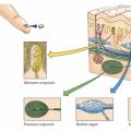 Types of skin receptors Krause flasks perceive