Somatosensory system. Tactile sensations
There are four types of skin sensations: tactile (sensation of touch, pressure), heat, cold and pain.
The sensation of touch is different from the sensation of pressure, for example the tongue cannot detect the pulse.
The number of tactile receptors is about 500,000, cold - 250,000, heat - 30,000. Most of the tactile receptors are located on the tips of the fingers, the palmar surface of the hand, the soles of the feet, tongue, border of the lower lip.
Touch stimulates rapidly adapting receptors, and pressure stimulates slowly adapting receptors. The fingertips and palm are especially sensitive to vibration. Tactile receptors, or mechanoreceptors, also respond to temperature stimuli. There are many temperature receptors on the face, especially on the lips and eyelids. Heat receptors are located deeper than cold ones, on the periphery of the cornea, and they are absent in the conjunctiva of the eye.
Until now, the relationship between the structure of skin receptors and their function has not been established. It is possible that the difference in sensations depends not only on the stimulation of different receptors, but on the peculiarities of the spatial and temporal distribution of impulses in the afferent nerve fibers and the speed of their conduction during stimuli of different quality and intensity (Neif, 1927). It is assumed that free nerve endings are organs of pain. Nerve fibers often end not between cells, but inside the cellular cytoplasm itself. This is an important fact, since any damaging agent that causes cell destruction or is an adequate stimulus for pain.
There is an opinion about the unity of peripheral receptors and peripheral nerve pathways for pain and tactile sensations. Irritation of the receptors of tactile sensations during subthreshold (for pain) and threshold stimuli causes "sub-pain" tactile sensations, which, when irritation intensifies, turn into a sensation of pain.
There are, however, classic examples of special pain reception: such are the pains arising from irritations of the cornea and eyelids, as well as from irritations of the celiac nerve, which do not give any other sensations. It is assumed that the receptors for the perception of heat and cold are the same. They differ only in the depth of their location in the thickness of the skin. Cold receptors are located more superficially.
The existence of four separate types of receptors in the skin analyzer is currently being questioned. The accuracy of counting skin receptors is also very relative, especially if we take into account the "watch" of skin receptors discovered in the laboratories of N. A. Rozhansky and L. A. Orbeli, which consists in their alternate excitation, which is revealed by the appearance of motor reflexes when individual skin points are irritated. The moment one of the skin receptors is excitable, the other is not. And the next moment, on the contrary, the first becomes excitable, and the second becomes excitable. This "watch" may be due to a change in excitation and inhibition in the neurons of the skin analyzer.
The excitability of the skin analyzer reaches a maximum by the age of 17-27 and changes dramatically depending on the functional state of the brain. For example, it is sharply reduced with fatigue and with strong emotions.
Simultaneous irritation of other analyzers (vision, hearing, smell, taste) also significantly reduces the excitability of the skin analyzer. Even moderate pain can be significantly reduced by simultaneous stimulation of other analyzers.
Absolute threshold irritation of tactile receptors is not the same in different parts of the body, the least - on the tongue and nose.
The excitability of tactile receptors is greatest at frequencies of mechanical displacements, or vibrations, 40 - 500 Hz. The accuracy of estimating the frequency of vibrations converted into nerve impulses reaches 5-10%.
Discrimination threshold (difference) about 1/30 (see p. 578).
Time threshold, i.e., the shortest time interval between two successively distinguishable stimuli is the smallest for a tactile analyzer (about 2 ms).
Consequently, the tactile analyzer is the most functionally mobile, or labile, followed by cold, heat and, finally, pain. The pain analyzer has the least functional mobility, individual pain stimuli differ to the least degree sequentially in time.
Simultaneous spatial threshold - the smallest distance between two points, at which they are felt separately with simultaneous stimulation, is different for all four types of skin reception, it is the smallest for tactile and the largest for pain reception.
The ability to combine tactile sensations received from different receptive fields into one complex sensation is developed throughout life due to the formation of temporary neural connections in the cerebral hemispheres. For example, touching a ball with the lateral surfaces of the fingers gives one sensation, and when crossing the fingers, two sensations of two balls are obtained (Aristotle's experiment).
Tactile irritations are very finely localized. This ability is developed throughout life. In addition to tactile receptors, it involves, in addition to tactile receptors, irritation of vision receptors, proprioceptors, etc. Regarding the threshold of tactile stimuli, it should be noted that it increases with age. Consequently, in the elderly, the ability to localize tactile irritation decreases.
Pain irritation can be localized to the least extent. In addition, severe pain is accompanied by irradiation of excitation in the central nervous system, which makes it impossible to localize it.
Adaptation in the skin analyzer
The skin analyzer is adaptable. Rapid adaptation to irritation leads to the fact that we do not feel the pressure itself, but only changes in pressure. When registering potentials in afferent nerves carrying impulses from tactile receptors, it is found that with continuous pressure on these receptors only during the first seconds the frequency of impulses reaches 250-350 per 1 s, and then it sharply decreases or the impulses stop, which is expressed in a decrease in the intensity of sensation. When the hand is immersed in warm water, we experience heat only for a short time, and then the skin analyzer adapts to temperature irritations, and the heat is not felt. When we change from warm to water at a lower temperature, we experience cold for a short time, and then become indifferent. Registration of potentials reveals a decrease in the frequency of afferent impulses or their termination. There is also adaptation to painful irritations. The injection into the skin is felt only for a short time, and then the sensation of pain stops, although the needle remains in the skin. The slower and the stronger the pain stimulation, the longer the flow of afferent impulses and, therefore, the slower the adaptation to pain.
It is assumed that in response to irritation of pain receptors, the oxidation of glucose and other substances in neurons involved in the pain reflex is accelerated. This leads to a lack of oxygen in them, which stops the conduction of pain impulses and causes natural inhibition of pain.
There are consistent sensations during stimulation of the tactile, temperature and pain receptors of the skin. After the end of the irritation of these receptors, tactile, temperature and pain sensations continue, then they disappear and after a while reappear. This wave-like attenuation and restoration of skin sensations is due to the wave-like nature of the nervous process in the skin analyzer. Conditioned reflexes are formed on skin irritation. Under the action of conditioned thermal stimuli of the skin, inhibition quickly occurs.
Pain problem, protopathic and epicritical sensitivity
Pain is of particular importance for life preservation. Pain is an indicator of a violation of normal processes, a danger signal that causes special protective reactions (contractions of the striated muscles, shifts in breathing, blood circulation, etc.), ensuring the safety of a given individual and species.
However, a strong (damaging), as well as a long-term effect of the "painful" stimulus, chronic irritation of pain analyzers turn the body's defense reaction into a harmful one, which is the cause of secondary disturbances of physiological processes.
Therefore, it is practically extremely important to develop techniques for stopping pain signaling by turning off receptors or afferent nerves and pathways, which leads to the elimination of pain.
Excitation of the caudate nucleus suppresses pain.
Pain is also caused by the humoral route - the appearance in histamine, substance P, serotonin, kinins and others (in fractions of μg). All these substances suppress interstitial respiration. There are kininogens in the blood, which, under the action of special enzymes, are converted into kinins - complex compounds of amino acids, for example, when contact factor XII appears during blood clotting. Histamine is formed from the amino acid histidine and, like other pain-causing substances, is destroyed very quickly.
Distinguish protopathic - pain and gross temperature sensitivity - and epicritical - tactile and subtle temperature sensitivity. Protopathic sensitivity is phylogenetically older and is inhibited by a phylogenetically younger epicritical sensitivity (Ged). This division of skin sensitivity is insufficiently substantiated.
The somatosensory system is a system of skin and musculoskeletal (proprioceptive) sensitivity, which provides the formation of tactile, temperature, pain and feelings of body position in space and movement of the structures of the musculoskeletal system.
The skin sensitivity system forms tactile, temperature and pain (nociceptive) sensations.
Tactile reception. mechanoreceptors
Tactile reception provides the sensation of touch, pressure, vibration, tickling, and itching, and is mediated by mechanoreceptors (Fig. 7.6). Mechanoreceptors of the skin, for their morphologically functional properties, are classified as primary receptors. They are exteroreceptors, therefore they transmit information about contact with the external environment.
Tactile receptors are divided into two groups:
■ encapsulated nerve endings or corpuscular receptors;
■ free nerve endings.
Encapsulated skin mechanoreceptors
Giachini Taurus - located in the skin and subcutaneous tissue, especially in the skin of the fingers, external genitalia, breast. They have an oval-shaped auxiliary structure of concentric layers of cells that surrounds the nerve ending of the afferent nerve fiber. Deformation of the auxiliary structure leads to the emergence of a receptor potential in the nerve ending. Receptors quickly adapt, transmit information by type Αβ nerve fibers (at a speed of 40-70 ms / s) about gross deformation of the skin and high-frequency vibrations.
Pacini corpuscles are irritated by rapid movement of tissues, therefore, they are important for assessing rapid mechanical effects. They are found at the junction of muscles and tendons, in the tissues of the joints.
FIG. 7.6.
Meissner Taurus - situated in hairless skin: finger kipe and feet, on the palmar and plantar surfaces, on the lips, centuries, external genitals, nipples of the mammary glands. They are placed on the edge of the epidermis and dermis in papillary layer dermis. Quickly adapt and transmit information nervous type типаβ fibers (speed 40 m / s) on the movement of light objects on the surface skin and low-frequency vibration.
Merkel discs - located in deep layers epidermis on palms and sole, slow adapt, transmit information by nerve fibers of type Αβ about skin touch, surface structure and precise localization of irritation.
Taurus Ruffini - located in the deep layers of the dermis and subcutaneous tissue, numerous in the plantar surface of the foot. They slowly adapt, transmit information nerve fibers such as Αβ about pressing.
Free nerve endings
Free nerve endings are located in the epidermis between epithelial cells. In the papillary dermis - located parallel to the dermo-elidermal limit. They quickly adapt, transmit information with Αδ nerve fibers (at a speed of 10-15 m / s) about pressure and touching the skin. The sensation of itching, tickling also occurs when free nerve endings located superficially in the skin are irritated, but information from them is transmitted by type C nerve fibers (speed 0.5-3 m / s).
Hair receptors belong to free nerve endings that surround hair follicles, react to hair displacement, adapt quickly. Information from them is transmitted by type Αβ nerve fibers.
From tactile mechanoreceptors, information is transmitted by afferent fibers in the central nervous system from the trunk and extremities to the spinal cord, from the head as part of the cranial nerves.
Assessment thresholds of tactile sensation occurs using the Frey esthesiometer, which allows you to determine the force of pressure arising on skin surface. The sensation threshold for different skin areas is different and is 50 mg for the most sensitive and 10 g for the least sensitive. Spatial resolution thresholds for tactile sensitivity allows you to estimate the density of receptors. they are determined using a Weber compass, has two "legs" with needles. Moving them apart, you can find the minimum distance at which two touches are perceived separately. This will be the spatial threshold of discrimination. For the receptors of the skin of the lips, it is 1 mm, for the skin of the pads of the fingers - 2.2 mm, for the skin of the hand - 3.1 mm, for the skin of the forearm - 40.5 mm, for the skin of the occiput and back - 54-60 mm, hips - 67.6 mm. Assessment of tactile sensation is important for the clinical picture of nervous diseases in the diagnosis of a lesion. various departments of the central nervous system.
Pathways. Medial Lemnisc System
Afferent nerves myelinated fibers from tactile mechanoreceptors pass to the spinal cord through rear roots and rise in the posterior columns in the medulla oblongata, where they form synapses with the neurons of the nuclei of the columns - thin / n. Gracilis) and wedge-shaped (n. Cuneatus) (Figure 7.7). This is where the second neurons begin and on the opposite side - intersect, forming a medial loop (lemniscus medialis), to which nerve fibers from the nuclei of the V pair of cranial nerves join. Then they ascend bilaterally to specific sensory switching nuclei of the thalamus. In the thalamus, the second neurons of the medial lemniscus system form synapses with the neurons of the ventral posterolateral nuclei (ventrobasal complex). Third thalamic neurons transmit information to the sensory cortex - the posterior central gyrus.
The lemnisc system ensures the transmission of information from mechanoreceptors, allows you to accurately localize the action of an irritating stimulus, to show the strength and gradation of the strength of sensation.

FIG. 7.7.
A feature of the lemniscus system is the spatial orientation of nerve fibers from individual parts of the body: fibers from the lower parts of the body lie in the posterior columns closer to the center, and from the upper parts - laterally.
In the nuclei of the thalamus, the head and face are presented medially in the ventrobasal complex of the nuclei, and the distal parts of the body - laterally.
Tactile information is also transmitted by the ventral spinothalamic tract from tactile receptors, information enters the posterior horns of the spinal cord, where it switches to the second neuron. The axons of the second neurons cross the segments of the spinal cord and pass into the anterolateral quadrant of the spinal cord to specific nuclei of the thalamus, where they switch to third neurons that carry information in the sensory cortex of the brain to the gelatinous substance of the posterior horns - plates I, II, III, they switch through 1-2 synapses to neurons, the axons of which cross the midline and move to the opposite side, where they rise upward as part of the anterolateral cord - anterolateral sensory system which includes ventral and lateral spinothalamic tract (Figure 7.8). Information from thermoreceptors goes mainly within the lateral spinothalamic tract to the ventrobasal complex of specific thalamic nuclei, and then to the sensory cortex. In addition, information is transmitted in the reticular formation of the brain stem and further through the nonspecific nuclei of the thalamus to the cerebral cortex. Sensory spinothalamic tract from thermoreceptors passing through specific kernels thalamus, called neospinothalamic tract .
The somatic sensory system provides a sensation that is generated by information from receptors in the body. These receptors can be classified into the following groups:
Mechanoreceptors, including tactile and proprioceptive;
Thermoreceptors (cold and heat)
Pain receptors that are activated when exposed to damage.
Characterization of tactile receptors. The sensations that arise when these receptors are excited - touch, pressure, vibration, perspiration, itching. Tactile receptors are located in different areas of the skin (epidermis and dermis). The sensation arises when the surface of the skin is irritated, and the pressure is deeper.
Tactile receptors there are 6 types:
1. Free nerve endings - polysensory, which can be excited by the action of both mechanical and thermal influences.
2. Meissner's little bodies - touch receptors, are encapsulated nerve endings. They adapt quickly. there are many of them on the skin of the fingers, palms, plantar surface.
3. Merkel discs - there are also many of them on the fingertips. Together with Meissner bodies, they participate in the localization of irritations. They are slow to adapt. Merkel discs are sometimes grouped into domed Pincus-Iggo receptors.
4. Rufin's little bodies - branched encapsulated endings of nerve fibers. They are located in the deep layers of the skin, poorly adaptable.
5. Taurus Pacini - The largest are large receptors that are bulbous. They are located deeper and in fascial tissues (Fig. 12.1). Pacini corpuscles are irritated by rapid movement of tissues, therefore, they are important for assessing rapid mechanical effects. They adapt quickly. They are found at the junction of muscles and tendons in the tissues of the joints, their size is from 0.4 to 0.5 mm.
6. Hair follicle receptors, formed by nerve fibers located at the base of the hair. They adapt quickly.
Characterization of tactile receptors
The sensations that arise when these receptors are excited are touch, pressure, vibration, perspiration, itching. Tactile receptors are located in different areas of the skin (epidermis and dermis). The sensation occurs when the surface of the skin is irritated, and the pressure is in the deep.
All tactile receptors are involved in determining the sensation of tissue vibration. At different frequencies of vibration, different receptors are excited. The sensation of tickling and itching is mainly associated with free nerve endings, they quickly adapt. Such receptors are found only in the surface layers of the skin. Itching is very important for recognizing insect crawling on the skin or a mosquito bite that causes itching.
Assessment of the thresholds of tactile sensation occurs with the Frey esthesiometer, which allows you to determine the force of pressure that occurs on the surface of the skin. The sensation threshold for different skin areas is different and is 50 mg for the most sensitive and 10 g for the least sensitive. Spatial resolution thresholds for tactile sensitivity provide an estimate of the receptor density. they are determined using a Weber compass, has two "legs" with needles. Pushing them apart, you can find the minimum distance at which two dots
Figure: 12.1. Scheme of the structure of skin mechanoreceptors in areas without hair (A) and with hair (B):
1 - stratum corneum, 2 - epidermis, 3 - corium, 4 - subcutaneous tissue, 5 - Meissner's little body, 6 - Merkel's disc, 7 - Pacini's little body, 8 - hair follicle receptor, 9 - tactile disc, 10 - Rufin's end
ki are perceived separately. This will be spatial discrimination threshold. For the receptors of the skin of the lips, it is 1 mm, for the skin of the pads of the fingers - 2.2 mm, for the skin of the hand - 3.1 mm, for the skin of the forearm - 40.5 mm, and for the skin of the occiput and back - 54-60 mm , hips - 67.6 mm.
Evaluation of tactile sensation is important for the clinical picture of nervous diseases when making a diagnosis of the impression of various parts of the central nervous system.
characteristic of proprioceptors
Proprioception provides the perception of posture and movements of our body. It provides deep, kinesthetic sensitivity. Proprioceptors - mechanoreceptors that irritate when stretched
Proprioceptors are divided into 2 groups:
1) muscle spindles;
2) the Golgi tendon organs.
Muscle spindles are in the muscles. They are attached to the working muscles in parallel, therefore, they are excited either when the extrafusal muscles are stretched, or when the muscle fibers of the spindles - intrafusal muscles contract. Because of this, they are called stretch receptors. These receptors are involved in the regulation of muscle length and in assessing the rate of change in muscle length.
Golgi tendon organs located in tendons, ligaments, joints. They are attached from one end to the muscle, and from the other to its tendons, therefore they are sequentially located in relation to the muscle, but they are also irritated by stretching, which occurs when the working muscle contracts and its tension increases. They are involved in the regulation of muscle tone.
thermoreceptor characteristic
Thermoreceptors are located not only in the skin, but also in internal organs and even in the central nervous system (hypothalamus). They are the primary receptors, as they are formed by free nerve endings and are divided into cold and heat.
The importance of thermoreceptors is not only in determining the temperature of the environment or objects. They play an important role in regulating the constancy of body temperature in humans and animals. Thermoreceptors are highly adaptable.
The concept of thermoreceptors is controversial. It is believed that the thermoreceptors in the skin are free nerve endings, as well as Ruffini's little bodies and Krause's flasks. It is believed that instead of the term "thermoreceptors" the term "hot spots" should be used, which are selectively sensitive to heat or cold. The lack of consensus is due to the fact that morphologically identifying heat or cold receptors was rather difficult. Before histological examination, tissues are frozen to make thin layer-by-layer sections, and it is not possible to establish the type of receptors that are sensitive to heat or cold. Taking this into account, it is advisable to use the term "thermal sensor", and the question of morphological identification remains for the future.
There is evidence that the number of temperature receptors (points) on the human skin is unstable and in the same area changes depending on the temperature of this area and a number of other factors. The lower the temperature of the skin and the environment, the more cold receptors and the less functional activity of the thermal ones. At high temperatures, the situation is the opposite. The hardening of the body is also important. In adapted people, the number of cold receptors in the cold is less than in unadapted people.
Wired and cortical parts of the somatic sensory system
From proprioceptors, impulses go as part of the afferent fibers of the A-alpha group (70-120 m / s), from tactile receptors - as part of the afferent fibers of the A-beta group (40-70 m / s) and A-delta (15-40 m / s), and for impulses coming from receptors that cause itching - as part of c-fibers (0.5-3 m / s). Conduction of impulses from thermoreceptors is carried out by fibers of the A-delta group and C-fibers.
From the trunk and limbs, impulses go as part of the spinal nerves, and from the head - as part of the trigeminal nerve. To conduct impulses that provide tactile sensitivity, the spinal-cortical tracts of Gaulle and Burdach are used.
Cortex representation of the somatic sensory system located in the postcentral gyrus cm-I (Fig. 12.2).
The cork representation of the somatosensory system is characterized by a number of features.
1. somatotopic organization - a certain location of the projections of body parts in it. The body is projected upside down in the postcentral gyrus.
2. The discrepancy between the sizes of these projections: the very territories are occupied by the tongue, lips, larynx, hand, as the most important for the assessment of irritation. Small areas - trunk and lower limb projections.
3. contralateral location of the projections. From the receptors on the left side, impulses go to the right hemisphere, and from the right side to the left hemisphere.
4. Consists mainly of monosensory neurons.
Irritation of the sm-I site leads to sensations identical to those that arise when exposed to stimuli (touch, vibration, heat, cold, rarely pain).
The associative site Cm-II is located at the lateral end of the postcentral gyrus on the superior wall of the Sylvian sulcus and consists mainly of polysensory neurons. It has a bilateral somatotopic representation of the body, therefore it plays an essential role in sensory and motor coordination of the two sides of the body (for example, when exposed to both hands).
Damage to the sm-I site - leads to a violation of the fine localization of sensations, and damage to the sm-II site - to astereognosia - unrecognizability of objects when feeling (without vision control).
The skin contains a variety of poorly differentiated receptors, which are divided: 1) into tactile ones, the irritation of which causes sensations of touch and pressure; 2) thermoreceptors - heat and cold; 3) painful.
Absolute specificity, i.e. the ability to respond to only one type of irritation is characteristic only of some receptor formations of the skin. Many of them react to stimuli of different modality. The emergence of various sensations depends not only on which receptor formation of the skin has been irritated, but also on the nature of the impulses coming from this receptor to the central nervous system.
Perception of mechanical stimuli (touch, pressure, vibration, stretching) is called tactile reception... Tactile receptors are found on the surface of the skin and mucous membranes of the mouth and nose. They are aroused by touching or pressing on them.
Tactile receptors include Meissner's bodies and Merkel discs, which are abundant on the fingertips and lips. Pressure receptors include Pacini's little bodies, which are concentrated in the deep layers of the skin, in the tendons, ligaments, peritoneum, and mesentery of the intestine. Nerve impulses originating in tactile receptors travel through sensory fibers to the posterior central gyrus of the cerebral cortex.
In different places of the skin, tactile sensitivity is manifested in different degrees. It is highest on the surface of the lips, nose, and on the back, sole, stand, abdomen is less pronounced. It is shown that simultaneous touching of two points of the skin is not always accompanied by the appearance of a sensation of two influences. If these points lie very close to each other, then there is a sensation of one touch. The smallest distance between points of the skin, when irritated, there is a sensation of two touches, is called threshold of space. The thresholds of space are not the same in different places of the skin: they are minimal on the tips of the fingers, lips and tongue and maximal on the hip, shoulder, and back.
Ambient temperature is exciting thermoreceptorsconcentrated in the skin, on the cornea of \u200b\u200bthe eye, in the mucous membranes. A change in the temperature of the internal environment of the body leads to the excitation of temperature receptors located in the hypothalamus.
Temperature receptors are very important in maintaining a constant body temperature, without which the vital activity of our body would be impossible.
There are two types of temperature receptors: cold and warm. Heat receptors are represented by Ruffini's bodies, cold receptors - by Krause cones. The bare endings of afferent nerve fibers can also function as cold and heat receptors.
Thermoreceptors in the skin are located at different depths: cold receptors are closer to the surface, heat receptors are deeper. As a result, the reaction time to cold stimuli is shorter than to heat. Thermoreceptors are grouped at certain points on the surface of the human body, while there are much more cold spots than heat spots. The severity of the feeling of warmth and cold depends on the place of the applied irritation, the size of the irritated surface and the ambient temperature.
Painful sensations arise with the action of any stimuli of excessive strength. The sensation of pain is of great importance for preserving life as a signal of danger, causing defensive reflexes of skeletal muscles and internal organs. However, damaging or prolonged irritation of pain receptors distorts the defensive reflexes, making them maladaptive.
The pain is localized less than other types of skin sensitivity, since the excitement arising from irritation of pain receptors spreads widely throughout the nervous system. Painful sensations also arise when a critical level of irritation of tactile receptors and thermoreceptors is reached. Simultaneous stimulation of the receptors for vision, hearing, smell and taste reduces the sensation of pain.
It is assumed that the onset of pain is associated with irritation of the endings of special nerve fibers. The obtained data indicate that the formation of histamine in the nerve endings is important in the formation of pain. The onset of pain is also associated with other substances formed in the tissues at the site of injury - bradykinin, factor XII of blood coagulation (Hageman factor), etc.
Pathways and cortical end of the skin analyzer. Excitation from the receptors of the skin analyzer is directed to the central nervous system through fibers having different diameters. Fibers of small diameter (with a conduction velocity of 30 m / s) switch to a second neuron in the spinal cord. The axons of these neurons as part of the anterior and lateral ascending pathways are directed, partially crossing, to the visual hillocks, where the third neuron of the cutaneous sensitivity path is located. The processes of these neurons reach the somatosensory zone of the pre- and postcentral gyrus of the cortex.
Thicker fibers (with a conduction speed of 30 to 80 m / s) pass without interruption to the medulla oblongata, where they switch to the second neuron. There is also a transfer to the second neuron of excitation coming from the receptors of the scalp. The axons of the neurons of the medulla oblongata are completely crossed at the level of the medulla oblongata and are directed to the visual hillocks. Excitation is transmitted to the somatosensory area of \u200b\u200bthe cortex along the axons of the neurons of the visual hillocks.
In the visual hillock, the skin surface of the head and face is represented in the posteromedial zone of the posterior ventral nucleus, and the upper and lower extremities, and the trunk, in its anterolateral part. There is a certain organization in the vertical arrangement of neurons that receive information from various parts of the skin surface. Above all, there are neurons that receive information from the skin surface of the legs, somewhat lower - from the body and even lower - from the arms, neck, head. The same arrangement is typical for the cortical section of the skin analyzer. Neurons that transmit information from the skin surface are divided into mono-, di- and polymodal. Monomodal neurons perform the function of discrimination, and di- and polymodal ones - integrative.
Age features of the skin analyzer. At the 8th week of intrauterine development, bundles of myelin-free nerve fibers are revealed in the skin, which end freely in it. At this time, a motor reaction appears to touch the skin in the mouth area. At the 3rd month of development, receptors of the lamellar body type appear. First of all, the nerve elements of the skin analyzer appear in the skin of the lips, then in the pads of the fingers and toes, then in the skin of the forehead, cheeks, and nose. Then, almost simultaneously, receptors are formed in the skin of the neck, chest, nipple, shoulder, forearm, armpit.
The early development of receptor formations in the skin of the lips provides the occurrence of the sucking act under the action of tactile stimuli. In the 6th month of intrauterine development, the sucking reflex is dominant in relation to the various movements of the fetus carried out at this time. It entails the emergence of various facial movements.
In a newborn, the skin is abundantly supplied with receptor formations and the nature of their distribution but its surface is the same as in an adult. In newborns and infants, the skin is most sensitive to touch in the mouth, eyes, forehead, palms of the hands and soles of the feet. The skin of the forearm and lower leg is less sensitive, and the skin of the shoulders, abdomen, back and thighs is even less sensitive - it corresponds to the tactile sensitivity of the skin of adults.
Newborns react to cold and warmth after a much longer period than adults, and to cold more than to heat, the skin of the face is most sensitive to heat. The sensation of pain in newborns is presented without localization of its source. The skin of the face is most sensitive to painful irritations. Localization of pain caused by irritation of interoreceptors is absent even in children 2-3 years old. There is no precise localization of all skin irritations in the first months or in the first year of life. By the end of the first year of life, children can easily distinguish between mechanical and thermal stimuli.
An intense increase in encapsulated receptors occurs in the first years after birth. At the same time, their number increases especially strongly in areas subject to pressure: for example, with the beginning of walking, the number of receptors on the plantar surface of the leg increases.
Since the hand acquires more and more importance in human life with age, the role of its receptor formations in the analysis and assessment of objects of the surrounding world, in the assessment of the movements carried out, increases. On the palmar surface of the hand and fingers, the number of polyaxone receptors increases, which are characterized by the fact that many fibers grow into one flask. In this case, one receptor formation transmits information to the central nervous system along many afferent pathways and, therefore, has a large area of \u200b\u200brepresentation in the cortex. An increase in the number of skin receptors can also occur in an adult, for example, after loss of vision and the need to navigate by touch.
During the first year of life, qualitative transformations of skin receptors take place, and only by the end of it all receptor formations of the skin approach in their morphological and functional characteristics to the adult state. Over the years, the excitability of tactile receptors increases, especially in the prepubertal and pubertal periods and reaches a maximum by the age of 17-25. During life, temporary connections of the musculocutaneous sensitivity zone with other perceiving zones are formed, which make it possible to clearly localize the resulting skin irritation.
Mental fatigue leads to a sharp decrease in the tactile sensitivity of the skin; for example, after five general education lessons, it can be halved. Exercise can increase skin sensitivity.
Human skin has tactile (touch), temperature and pain receptors. Different types of receptors differ in their structure and are distributed in the skin in the form of a kind of mosaic.
Tactile receptors perceive mechanical stimuli, accompanied by a sense of touch and pressure. They are in the form of elongated bulbs, to which nerve endings fit. Tactile receptors include: tactile bodies (Meissner bodies) having the appearance of one tortuous nerve endings, dressed in a capsule; lamellar bodies (Pacini's little bodies), consisting of a nerve endings surrounded by connective tissue plates; tactile discs of Merkel, located near the hair follicles, in the epidermis, as well as in the vessels and in the deep layers of the skin on the surface of the hand, in the area of \u200b\u200bthe palms, as well as on the tips of the fingers, lips, tendons, peritoneum and mesentery of the intestine, etc. there are 25 tactile receptors per 1 cm2 of skin.
More receptors in the skin of the palms, at the ends of the fingers, on the lips and the tip of the tongue; least of all - in the skin of the back and abdomen. The threshold of irritation of the most sensitive areas is 50 mg, and in the least sensitive - up to 10 g. According to their functional characteristics, tactile receptors are divided into phase and static Phase tactile receptors are excited by dynamic stimulation, they have high sensitivity, a short latency period and quickly adapt. Static tactile receptors are excited mainly from static stimuli, they are less sensitive, but have a longer latency period and adapt more slowly.
Excitation that occurs in the tactile receptors upon contact of the skin with objects enters the cerebral center of the tactile analyzer, localized in the I region of the somato-sensory area of \u200b\u200bthe cerebral cortex (posterior central twist of the cerebral cortex), where it is transformed into a sensation of touch or pressure. The differentiation of this sensation depends on the adaptive abilities of the tactile receptors of the skin: As mentioned above, phasic tactile receptors are easily adapted and they respond only to changes in the intensity of the stimulus and give a short-term sensation of touch, even if the pressure stimulus acts for a long time. Static tactile receptors, adapt slowly, are excited only with prolonged exposure to a mechanical stimulus, which provides a feeling of pressure. By the touch mechanism, irritation in the form of vibration can also be perceived. Thanks to tactile sensitivity, a person feels the shape, size and nature of the surface of the surrounding objects. For contact is also characterized by a spatial sensation, which consists in the ability to distinguish and perceive as separate, two simultaneously irritated points of the body.
Thermoreceptors, or temperature receptors, are two types of nerve endings. Some of them (Krause cones) perceive mainly cold stimuli, and the second (Ruffini's bodies) perceive thermal stimuli. Placed thermoreceptors in the skin as well as in the mucous membrane of the nose, mouth, larynx, esophagus, stomach and intestines. In terms of structure, thermoreceptors are glomeruli of thin nerve endings that are contained in connective tissue capsules. irritates the skin's thermoreceptors and causes sensations of heat or cold in the analyzer's cork. As a result, the lumen of the blood vessels of the skin changes reflexively, thereby changing its blood supply and temperature.
There are about 250 thousand cold receptors in the body, up to 30 thousand heat receptors. Cold receptors are located at a depth of 0.17 mm, and heat receptors - 0.3 mm from the skin surface. Due to this, heat receptors are excited relatively slowly, while cold receptors react very quickly, both to irritation with a temperature below 18-20 ° C and to irritation with a temperature above 40-45 ° C (for example, the "goose bumps" effect when the body is immersed in hot water ). Thermoreceptors constantly inform the body about the state and changes in the ambient temperature and is the most important link in maintaining the temperature (thermostasis) of the body. In children, the feeling of temperature is manifested already from the first days after birth.
Pain is a specific feeling, qualitatively different from any other feeling. It arises when an irritant acts on one or another part of the body and has a destructive character. In this case, a whole line of protective reactions occurs, aimed at preserving parts of the body or the whole organism.
Painful irritations are perceived by pain receptors, or free nerve endings. Pain receptors are located not only in the skin, but also in muscles, bones, and internal organs. There are about 100 pain points on the surface of 1 cm2, and there are about a million of them on the entire surface of the skin. There is almost no area on the skin where there would be no pain receptors, but they are located unevenly: more in the axillary and inguinal areas and least of all on the soles, palms, ears. Excitation arising in the pain receptors as a result of the action of the stimulus are transmitted along the centripetal nerves to the higher cortical and subcortical (in the thalamus and hypothalamus) pain centers, where pain sensations are formed. The strength of painful sensations largely depends on the state of the nervous system. Pain receptors respond to significant fluctuations in temperature, pressure, and the concentration of prostaglandins secreted by damaged body cells. The penetration of information about the localization and intensity of pain into the centers of the brain stimulates the release of endorphins into the blood, which are pain blockers.
With painful irritations, the normal activity of the body is reflexively disrupted, and especially: the release of adrenaline into the blood increases, the concentration of sugar in the blood rises, the rhythm of heart contractions is disturbed, blood clotting accelerates, blood pressure rises, breath is held, etc. With very strong pain irritations, painful shock can be observed (temporary loss of consciousness, dizziness, fainting).
Another type of skin sensitivity is the sensation of tickling, which provides nerve endings freely located in the surface layers of the skin. This type of receptor is characterized by specific responses to stimuli of varying intensity. The tickling sensation is associated with the activation of this group of receptors, which gave the name to the receptors themselves - the tickling receptors.
Due to the action of thermal factors, chemicals, electric current or ionizing radiation, damage to body tissues and, above all, skin, called burns, can occur. Four degrees of burns are distinguished by the depth of tissue damage. Burns of the first degree are characterized by local (erythel), slight swelling and an increase in local temperature, lasts 2-5 days and usually passes without a trace. Second degree burns also cause local redness and swelling of the skin, and in addition, they are also characterized by the appearance of bubbles filled with yellowish liquid (lymph). These burns are accompanied by pain and fever.
Burns of III-A degree are accompanied by III-A degree necrosis of all layers of the skin, and IV degree - necrosis of the skin and deep tissues. Emergency care for burns consists in the immediate removal and neutralization of the factor that caused it. In case of chemical burns, the affected skin and mucous membranes should be immediately washed with plenty of cold running water (for at least 15 minutes). In the event of a skin burn with sulfuric acid or quicklime, it is impossible to rinse the affected area with water, since it will only enhance their effect. For this, oil or animal oil is used. In case of severe lesions, patients are hospitalized.
 Abraxas is destroying the Marvel Universe!
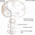 Somatosensory system. Tactile sensations. Classification and structure of receptor formations of the skin analyzer Where are the largest concentrations of tactile receptors
 Best congratulations Cool obscene birthday greetings from a friend